Essential lab manuals, like Seeley’s, guide students through practical exercises, fostering a deeper understanding of human anatomy and physiology, beginning January 15, 2026․
Purpose of a Lab Manual
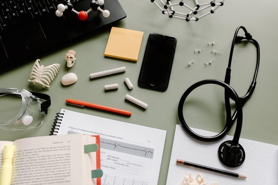
A lab manual serves as a crucial companion, detailing objectives, instructions, and content for practical anatomy and physiology exercises, like BIO201’s organization overview․ It enhances learning through hands-on application, supplementing textbook knowledge with direct observation and dissection, ensuring a comprehensive understanding of the human body․
Importance of Hands-on Learning
Hands-on learning is paramount in anatomy and physiology, moving beyond theoretical knowledge to practical application․ Lab manuals, like those accompanying Martini Art, facilitate dissection and specimen handling, solidifying concepts and developing critical thinking skills․ This active engagement fosters deeper retention and a more intuitive grasp of bodily structures․
Basic Anatomical Terminology
Understanding anatomical language – directional terms and body planes – is foundational for accurately describing human anatomy, as detailed in lab exercises and manuals․
Anatomical Position
The standard anatomical position – standing erect, feet slightly apart, palms facing forward – provides a universal reference point for describing body parts and their relationships․
Lab manuals emphasize this position for consistent orientation during dissections and observations, ensuring clear communication of anatomical structures and functions․
Accurate descriptions rely on this baseline, vital for understanding directional terms and body planes explored in introductory anatomy and physiology courses․
Directional Terms (Superior, Inferior, etc․)
Laboratory manuals thoroughly define directional terms – superior/inferior, anterior/posterior, medial/lateral – crucial for pinpointing anatomical locations accurately․
Understanding these terms, alongside proximal/distal, is fundamental for describing relationships between body structures during dissections and clinical observations․
Consistent application, as taught in manuals, ensures precise communication within the field of anatomy and physiology, starting February 17, 2026․
Body Planes (Sagittal, Frontal, Transverse)
Essential lab manuals detail body planes – sagittal, frontal (coronal), and transverse – vital for sectioning and visualizing anatomical structures in three dimensions․
Understanding these planes allows for accurate description of anatomical sections, crucial for imaging interpretation and surgical planning, beginning January 15, 2026․
Practical exercises within manuals reinforce spatial reasoning and anatomical orientation, enhancing comprehension of complex anatomical relationships․

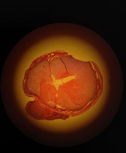
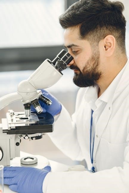
Microscopic Anatomy: Histology
Essential manuals introduce histology, the study of tissues, utilizing microscopy to examine epithelial, connective, and muscle tissue structures, starting January 15, 2026․
Epithelial Tissue Types
Essential laboratory exercises focus on identifying epithelial tissue types – squamous, cuboidal, and columnar – based on shape and layering, utilizing microscopic observation․
Students learn to differentiate between simple and stratified arrangements, understanding their roles in covering and lining body surfaces, starting January 15, 2026, with detailed manuals․
Connective Tissue Types
Essential lab work involves recognizing connective tissue varieties: connective tissue proper, cartilage, bone, and blood․ Students analyze their unique matrix compositions and cellular elements․
Manuals guide identification of dense and loose connective tissues, hyaline and elastic cartilage, and blood components, enhancing understanding of support and transport functions, beginning January 15, 2026․
Muscle Tissue Types
Essential laboratory exercises focus on distinguishing skeletal, smooth, and cardiac muscle tissues․ Students examine microscopic structures, noting striations and cellular arrangements․
Lab manuals aid in identifying muscle fiber characteristics and understanding their roles in movement and bodily functions, beginning January 15, 2026․ Practical application solidifies theoretical knowledge․
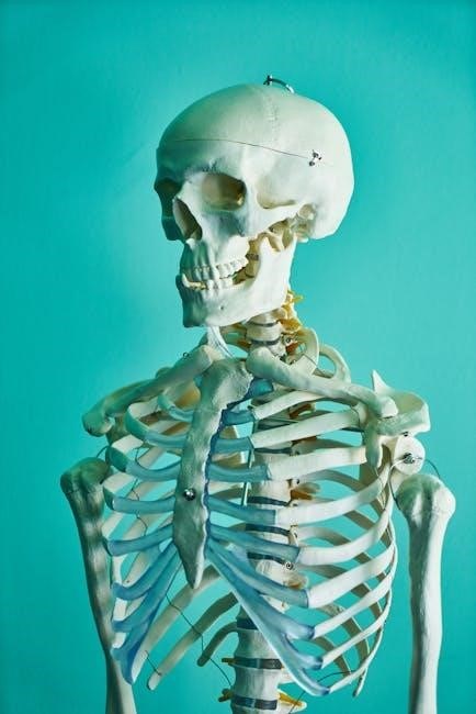
Skeletal System
Essential labs involve bone classification, identifying markings, and exploring joint structures․ Manuals facilitate understanding skeletal anatomy and its functional relationships, starting 02/17/2026․
Bone Classification
Essential laboratory exercises focus on categorizing bones – long, short, flat, irregular, and sesamoid – based on shape and function․ Manuals aid in identifying these types through observation and dissection․ Understanding classification is crucial for grasping skeletal anatomy, as detailed in lab resources available since January 15, 2026, and is vital for comprehending biomechanics and movement․
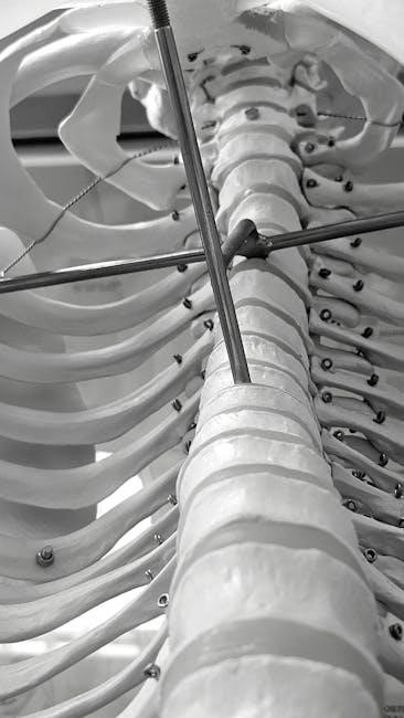
Bone Markings
Essential lab work involves identifying bone markings – projections, depressions, and openings – serving as muscle attachment sites or passageways for vessels and nerves․ Manuals guide students in recognizing features like tubercles, foramina, and fossae․ Accurate identification, starting January 15, 2026, is key to understanding skeletal function and anatomical relationships․
Joint Classifications
Essential laboratory exercises focus on classifying joints structurally (fibrous, cartilaginous, synovial) and functionally (synarthrotic, amphiarthrotic, diarthrotic)․ Lab manuals aid in differentiating these types, relating structure to movement․ Understanding joint classifications, beginning January 15, 2026, is crucial for analyzing skeletal system mechanics and range of motion․
Muscular System
Essential labs explore muscle fiber types, actions, and major muscle groups, enhancing comprehension of movement mechanics and physiological functions, starting January 15, 2026․
Muscle Fiber Types
Laboratory exercises focus on differentiating skeletal muscle fiber types – slow oxidative, fast oxidative glycolytic, and fast glycolytic – based on contraction speed and fatigue resistance․
Students will analyze histological slides and relate fiber type composition to specific muscle functions and athletic performance, beginning January 15, 2026, utilizing essential manuals․
Understanding these distinctions is crucial for comprehending muscular adaptations to training and individual physiological responses․
Muscle Actions
Lab activities explore muscle actions – agonist, antagonist, synergist – through movement analysis and dissection, enhancing comprehension of biomechanics․
Students will identify muscles responsible for specific movements, observing how they cooperate and oppose each other, starting January 15, 2026, with essential manuals․
Practical application reinforces understanding of how muscles work together to produce coordinated motion․
Major Muscle Groups
Laboratory exercises focus on identifying and palpating major muscle groups – limbs, trunk, head – using anatomical models and dissection techniques․
Students learn origins, insertions, and actions of key muscles, correlating structure with function, beginning January 15, 2026, with essential manuals․
Practical experience builds a foundational understanding of muscular anatomy and its role in human movement․
Nervous System
Lab manuals detail neuron structure, brain and spinal cord anatomy, emphasizing practical identification and functional relationships, starting February 17, 2026․
Neuron Structure and Function
Laboratory exercises focus on identifying neuron components – dendrites, cell body, axon – and understanding their roles in signal transmission․ Manuals explore action potentials, synapses, and neurotransmitter functions, crucial for nervous system comprehension․ These essential studies, beginning in 2026, build a foundation for advanced neurological concepts, utilizing practical dissection and observation techniques․
Brain Anatomy
Lab manuals guide dissection and identification of major brain regions – cerebrum, cerebellum, brainstem – and their associated functions․ Students explore lobes, fissures, and functional areas, enhancing understanding of neurological control․ These essential exercises, starting February 17, 2026, utilize models and diagrams for comprehensive anatomical study, building a strong neurological base․
Spinal Cord Anatomy
Laboratory manuals facilitate the study of the spinal cord’s structure, including gray and white matter, dorsal and ventral horns, and nerve roots․ Students trace pathways and identify key anatomical features, crucial for understanding reflex arcs and neural communication․ These essential exercises, beginning in 2026, build a foundation for neurological function․
Cardiovascular System
Lab manuals detail heart anatomy, blood vessel structures (arteries, veins, capillaries), and blood components, enabling students to trace circulatory pathways and understand function․
Heart Anatomy
Laboratory manuals facilitate dissection and identification of the heart’s chambers – atria and ventricles – alongside valves ensuring unidirectional blood flow․ Students explore the coronary arteries, vital for myocardial perfusion, and examine the pericardium’s protective layers․
Detailed diagrams and exercises within these manuals enhance comprehension of the heart’s complex structure and its crucial role in the cardiovascular system․
Blood Vessels (Arteries, Veins, Capillaries)
Lab manuals guide students in differentiating arteries, veins, and capillaries based on structural features like wall thickness and valve presence․ Dissection exercises and microscopic observations reveal how these vessels contribute to systemic and pulmonary circulation․
Understanding blood flow dynamics and vessel function is crucial, as highlighted in human anatomy and physiology studies․
Blood Components
Laboratory manuals facilitate the identification of blood components – erythrocytes, leukocytes, and platelets – through microscopic analysis of stained slides․ Hematocrit and blood typing exercises demonstrate key physiological functions․
Students learn about the roles of each component in oxygen transport, immunity, and coagulation, essential concepts in human anatomy and physiology․
Respiratory System
Lab manuals detail respiratory structures like lungs and the trachea, alongside pulmonary ventilation exercises, crucial for understanding gas exchange in human physiology․
Respiratory Structures (Lungs, Trachea, etc․)
Laboratory manuals meticulously guide dissection and identification of key respiratory structures, including the lungs, trachea, bronchi, and diaphragm․ These exercises emphasize anatomical relationships and functional roles․ Students learn to trace airflow pathways and observe histological differences within these structures, enhancing comprehension of human anatomy and physiology;
Pulmonary Ventilation
Lab manuals detail experiments exploring pulmonary ventilation mechanics, utilizing spirometers to measure vital capacity and tidal volume․ Students analyze data to understand factors influencing airflow, like resistance and lung compliance․ These practical applications solidify comprehension of human physiology, bridging theoretical knowledge with real-world physiological measurements․
Digestive System
Lab manuals explore digestive organs and functions through dissection and observation, clarifying processes like absorption and peristalsis for a comprehensive physiology understanding․
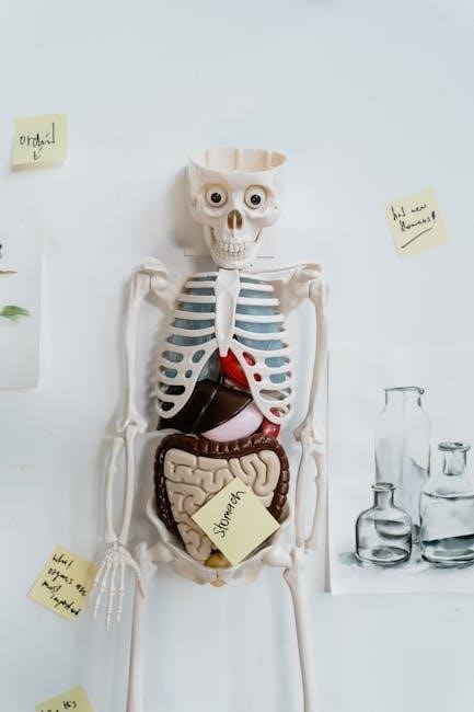
Digestive Organs and Functions
Laboratory exercises meticulously detail each organ – mouth, esophagus, stomach, intestines, liver, and pancreas – and their specific roles in breaking down food․ Manuals emphasize mechanical and chemical digestion, nutrient absorption, and waste elimination․ Students learn how structures correlate with functions, utilizing dissection and observation to understand the physiology of each component, enhancing comprehension of the entire digestive process․
Digestive Processes
Lab manuals guide students through investigations of key processes: ingestion, propulsion, mechanical breakdown, chemical digestion, absorption, and defecation․ Experiments demonstrate enzyme activity, peristalsis, and the impact of different food types․ Students analyze how these processes work in concert, utilizing models and simulations to visualize complex interactions within the digestive system, solidifying their understanding․
Urinary System
Lab manuals detail kidney anatomy and urine formation, utilizing models to explore nephron structure and function, crucial for understanding bodily fluid regulation․
Kidney Anatomy
Laboratory exercises focus on identifying kidney structures – cortex, medulla, pelvis, and associated vessels – through dissection and models․ Students learn to trace blood flow, examining nephrons and renal tubules․
Manuals emphasize the relationship between anatomical features and physiological processes, like filtration and reabsorption, vital for maintaining homeostasis․ Detailed diagrams and labeling activities enhance comprehension of this complex organ’s structure․
Urine Formation
Lab manuals detail the processes of glomerular filtration, tubular reabsorption, and tubular secretion, crucial for urine formation․ Experiments may involve analyzing urine samples to assess kidney function and identify abnormalities․
Students learn how hormonal controls, like ADH and aldosterone, influence water and electrolyte balance during urine production․ Visual aids and interactive exercises clarify these complex physiological mechanisms․

Endocrine System
Lab manuals explore hormone functions and major endocrine glands through experiments, examining their impact on body systems and physiological processes․
Major Endocrine Glands
Laboratory exercises focus on identifying and understanding the roles of key glands – pituitary, thyroid, adrenals, pancreas, and gonads – within the endocrine system․
Manuals detail their anatomical locations and hormonal secretions, emphasizing how these glands regulate vital functions like metabolism, growth, and reproduction, starting February 17, 2026․
Hormone Functions
Lab manuals explore how hormones, chemical messengers, impact target cells and tissues, influencing physiological processes․
Students investigate hormone mechanisms – including negative and positive feedback loops – and their effects on metabolism, growth, and reproduction, utilizing resources available since January 15, 2026․

Reproductive System
Lab manuals detail male and female reproductive anatomy, exploring gametogenesis and hormonal control, essential for understanding human biological functions․
Male Reproductive Anatomy
Laboratory manuals meticulously illustrate the male reproductive system, covering the testes, epididymis, vas deferens, seminal vesicles, prostate, and bulbourethral glands․
Detailed diagrams and exercises focus on sperm production (spermatogenesis), hormone regulation, and the anatomical pathways crucial for reproductive function, aiding comprehensive understanding․
Female Reproductive Anatomy
Lab manuals provide detailed explorations of female reproductive structures, including the ovaries, fallopian tubes, uterus, cervix, and vagina․
Exercises emphasize oogenesis, the menstrual cycle, hormonal control, and the anatomical changes during pregnancy, offering a thorough understanding of female reproductive physiology and anatomy․
Sensory Systems
Lab manuals dissect eye and ear anatomy, exploring sensory receptors and neural pathways crucial for vision and hearing, enhancing physiological understanding․
Eye Anatomy
Laboratory manuals facilitate detailed exploration of the eye’s intricate structures, including the cornea, lens, retina, and optic nerve․ Dissections and diagrams aid in visualizing light’s path and image formation․
Students learn to identify key anatomical features and understand their roles in visual perception, connecting structure to function within the sensory system․
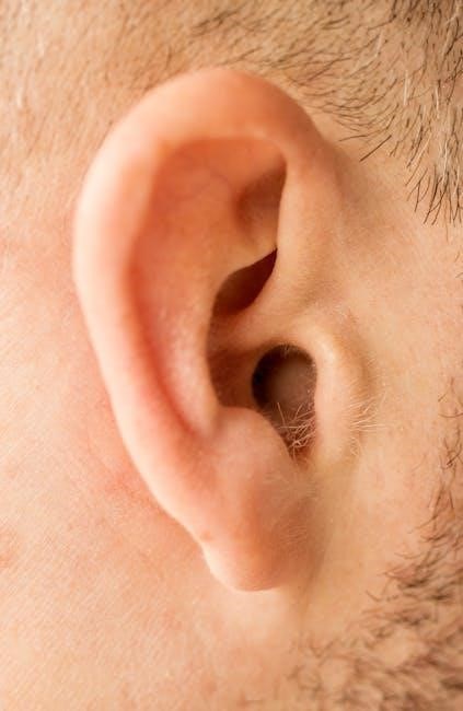
Ear Anatomy
Lab manuals provide comprehensive study of the ear’s anatomy – outer, middle, and inner ear – focusing on structures like the tympanic membrane, ossicles, and cochlea․
Exercises involve tracing sound wave transmission and understanding the mechanisms of hearing and balance, crucial for sensory system comprehension, as of February 17, 2026․
Laboratory Safety and Procedures
Manuals emphasize specimen handling, proper dissection techniques, and safety protocols, ensuring a secure learning environment for anatomy and physiology studies․
Proper Handling of Specimens
Laboratory manuals detail crucial guidelines for respectful and safe specimen management․ This includes appropriate personal protective equipment (PPE), careful observation, and meticulous documentation․
Proper preservation techniques, alongside responsible disposal methods, are also highlighted, ensuring both student safety and ethical considerations are prioritized throughout the anatomy and physiology coursework․
Dissection Techniques
Laboratory manuals provide detailed, step-by-step instructions for precise dissection․ These guides emphasize the correct use of instruments, careful tissue separation, and accurate identification of anatomical structures․
Students learn to approach dissections systematically, respecting the specimen while gaining invaluable hands-on experience in human anatomy and physiology, enhancing their understanding․